

Anything followed by ‘tear’ sounds awfully serious. But estimates are that by old age (clinically, 65) about two thirds of adults will show meniscus tears on MRI or CT. Strangely enough, of that two thirds, only some of them will have knee pain that is related to that tear – others will have no pain, or knee pain that is unrelated to the tear.
So what are meniscus tears and why do we care about them?
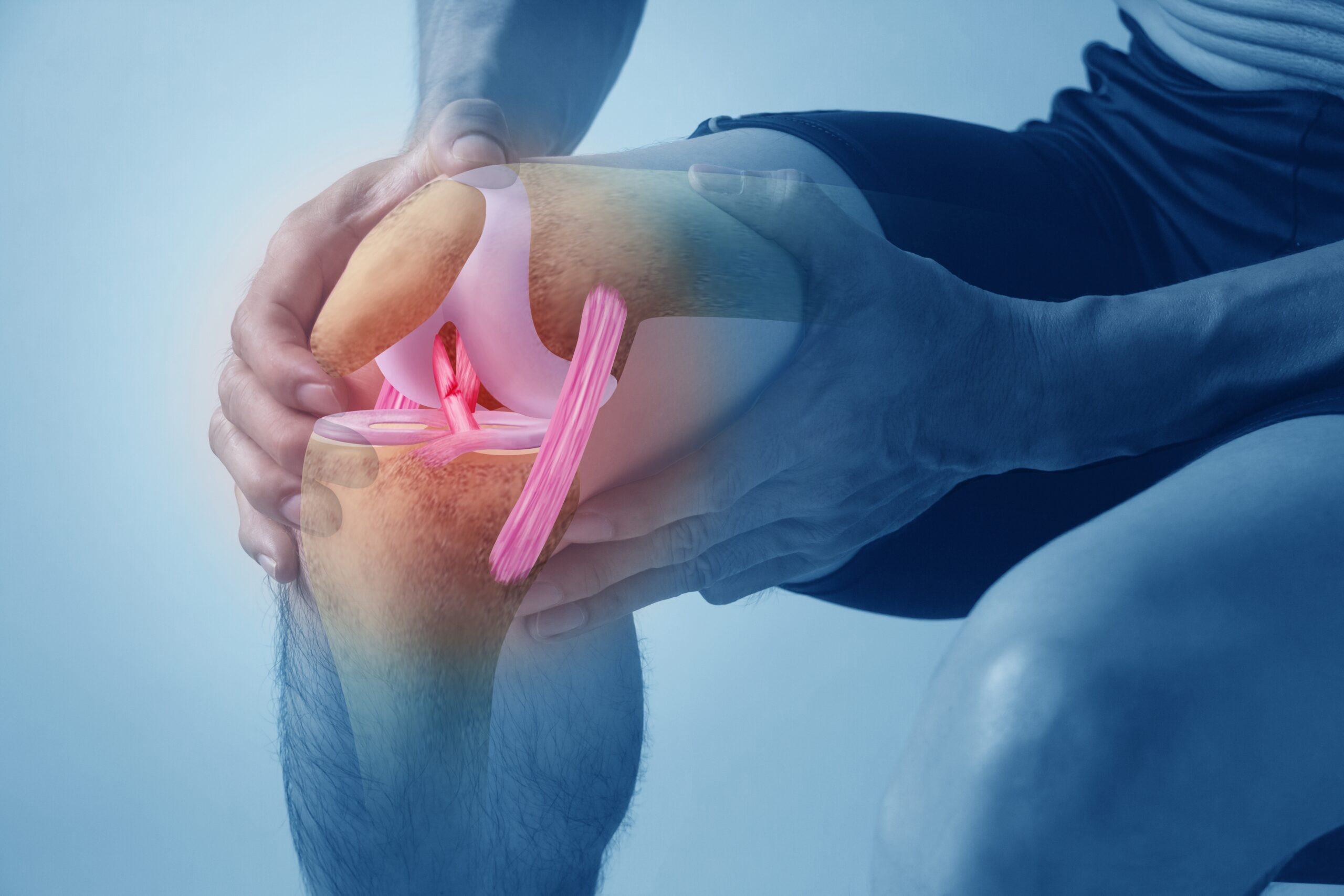
A meniscus in the knee is a thick, fibrocartilaginous structure which sits between the main bones of the knee joint and serves to absorb shock and smooth and stabilise knee joint movement. It doesn’t have a fabulous blood supply, in fact, only the very outer layers have a blood supply. This lack of blood supply means that damage done to a meniscus is unlikely to be repaired by your body, unless it’s really superficial – or, strangely, really deep.
Tears are often categorised by the shape and size when imaged. Radial and horizontal tears tend to be more common variants and occur with overload/sub-optimal loads of the knee along with age related changes. Bucket handle tears, where a portion of the meniscus has separated and formed a flap, tend to come from traumatic injury to the knee and are generally associated with greater knee dysfunction.
While the size and shape parameters are a useful tool to describe meniscus injuries, they do not adequately convey the whole picture. For instance, an image will not tell us why the injury occurred in the first place- what muscle imbalances or motor strategies led to the overloading of the knee? Did the individual land awkwardly?
In general, we don’t need an MRI or CT scan to tell you have a meniscus tear, there is often a characteristic clicking or locking sensation and there are a few tests your physiotherapist can perform to tell if you have a symptomatic meniscus tear. If you have severe dysfunction or are not responding to conservative management, your physiotherapist may choose to order an MRI and refer you on to an orthopaedic specialist.
What next?
For most acute, traumatic meniscus injuries, where there is not significant trauma to other structures in the knee, exercise based rehabilitation, is considered the first-line management strategy. A mix of bracing and taping may be used in the early phases along with retraining of movement strategies and strengthening in the later phases. At BEST this takes 4-6 months to return to sport. Some individuals may have significant damage and dysfunction that does not respond well to rehabilitation, in these cases your physiotherapist may refer you on to an orthopaedic specialist to consider surgery. Even if an injury progresses to surgical repair, your rehabilitation is not wasted, as pre-operative condition impacts how well and how quickly you recover.
Depending on the damage, surgeons may opt to repair and stabilise the tissue or remove parts of meniscus that are impeding movement. Post-operative rehabilitation is a non-negotiable.
More chronic meniscus injuries – those which have occurred over years of loading, will take longer to resolve. Resolve? But you said before the body doesn’t heal them very well!
The underlying structure does not often heal, but the function of the knee can be restored. This may take approximately 8-12 weeks to see improvements in function, but longer to achieve full resolution.
In either case, your physiotherapist will assess your underlying imbalances and movement strategies to normalise function as much as possible.